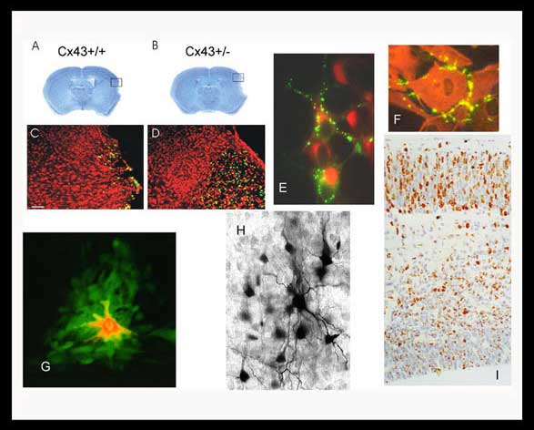
A,B,C,D: Decreased connexin43 expression results in increased infarct size and neuronal death (apoptosis green cells) following stroke injury in mice.
E: Connexin43 tagged with GFP enable us to visualize gap junction formation in live neurons.
F: Astrocytes in culture display localization of connexin43 at areas of intercellular contact.
G: A low molecular weight fluorescent dye (green) injected into a single cell (red) demonstrates the presence of gap junctions through the spread of the green dye.
H: Similar spread of a low molecular weight marker (neurobiotin) is also observed when a single neuron is injected in a brain slice.
I: The migration of new neurons can be followed as they are marked with bromodeoxyuridine in the developing cerebral cortex.


